Abstract
Disseminated tuberculosis with central nervous system involvement is difficult to diagnose because early microbiologic testing may be negative despite severe disease. This case highlights a 43-year-old immunocompetent man from Senegal who presented with headache, weight loss, night sweats, cough, miliary pulmonary nodules, and extrapulmonary lesions concerning for disseminated tuberculosis. Initial sputum and cerebrospinal fluid MTB-PCR were negative, but inflammatory cerebrospinal fluid findings supported tuberculous meningitis, prompting empiric anti-tubercular therapy with corticosteroids. This case shows that a negative initial PCR should not delay treatment when clinical suspicion is high and adequate repeat sampling may improve diagnostic yield.
Background
Tuberculosis (TB) is a chronic infectious disease caused by Mycobacterium tuberculosis that most commonly affects the lungs but can involve nearly any organ system. It remains a major global health burden, and extrapulmonary disease can present with variable, nonspecific features that delay recognition.1,2 While extrapulmonary TB is not uncommon, central nervous system involvement is comparatively rare, with tuberculous meningitis comprising roughly 1 to 5% of TB cases and most often seen in children and immunocompromised hosts rather than immunocompetent adult.3 We present a case of multi-organ disseminated TB with tuberculous meningitis in an immunocompetent adult, highlighting the diagnostic challenge posed by an initially negative CSF MTB-PCR and the importance of early clinical suspicion.
 Case Report
A 43-year-old male from Senegal with no prior medical history presented to the emergency department on the evening of admission with a two-day history of worsening headache. He reported abdominal pain, productive cough without hemoptysis, night sweats, cycling fevers, and a 15 kg weight loss over two months. He had relocated from Senegal two years prior with no subsequent travel. During the symptomatic period he had self-administered azithromycin and spiramycin from his home country without improvement. He denied tobacco and alcohol use, any known contact with active tuberculosis, and prior tuberculosis.
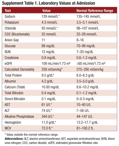
On admission he was afebrile (36.8°C) and tachycardic (115 beats per minute), with blood pressure 123/79 mmHg, respiratory rate 23, and oxygen saturation 95% on room air. Examination was notable for costovertebral angle tenderness at T11–T12, without palpable lymphadenopathy or organomegaly. Laboratory evaluation revealed microcytic anemia, transaminitis with markedly elevated alkaline phosphatase, and hyponatremia, as detailed in Table 1.
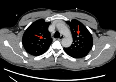
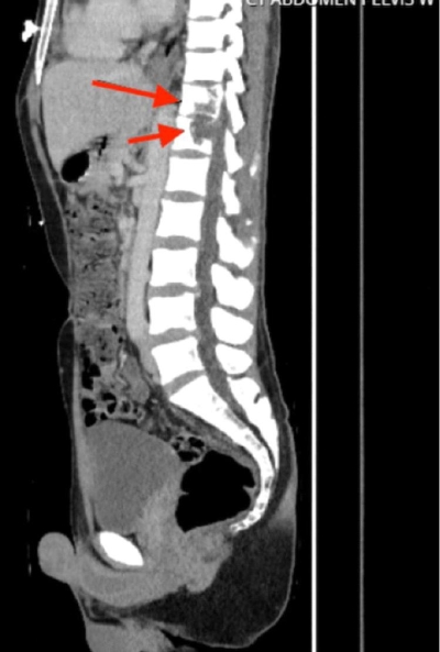
Cross-sectional imaging was obtained on the day of admission. Chest CT demonstrated innumerable 1 mm pulmonary nodules bilaterally consistent with a miliary pattern (Figure 1). CT of the thoracolumbar spine demonstrated destructive lytic changes at T11–T12 with intervertebral sparing, consistent with Pott’s disease (Figure 2). CT abdomen/pelvis revealed a 2.7 × 1.6 cm left prostatic hypodensity without rim enhancement. Dedicated MRI of the cervical, thoracic, and lumbar spine confirmed the T11–T12 involvement. The prostatic lesion was not biopsied and was therefore considered a radiographically suspected rather than microbiologically confirmed focus.
An infectious workup included acid-fast bacilli (AFB) culture, Quantiferon-TB Gold, sputum MTB-PCR, hepatitis panel, and HIV screening. Hepatitis B serologies obtained on hospital day 4 showed chronic active infection with an HBV DNA viral load of 20 IU/mL, and entecavir 0.5 mg orally daily was started the same day. Given the hepatotoxicity of both hepatitis B and several first-line anti-tubercular agents, liver enzymes were monitored throughout admission and remained only mildly elevated, allowing treatment to continue unchanged. Quantiferon-TB Gold was positive, while sputum MTB-PCR was negative, possibly reflecting reduced sensitivity after prior partial antimicrobial exposure.
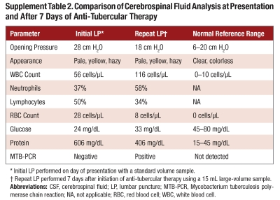
The following day (hospital day 1), the patient developed worsening headache and photophobia. Neurologic examination was grossly intact and notable for nuchal rigidity, with negative Kernig and Brudzinski signs. A first lumbar puncture was performed that evening. CSF analysis demonstrated an elevated opening pressure of 28 cm H2O and a mixed pleocytosis with 56 white blood cells per µL (37% neutrophils, 50% lymphocytes), markedly elevated protein at 606 mg/dL, and a profoundly low glucose of 24 mg/dL. Initial CSF MTB-PCR was negative.
Given the miliary nodules, multi-organ involvement, positive Quantiferon-TB Gold, inflammatory CSF profile, and epidemiologic background, empiric anti-tubercular therapy was initiated the same day, with oral isoniazid (300 mg daily), oral pyrazinamide (1,500 mg daily), oral ethambutol (1,200 mg daily), oral pyridoxine (50 mg daily), and intravenous rifampin (900 mg daily). Intravenous levofloxacin (750 mg daily) was added for its favorable CSF penetration, although fluoroquinolones are not standard first-line therapy for tuberculous meningitis and lack a consistent survival benefit in randomized trials. Adjunctive dexamethasone was administered to mitigate the inflammatory response associated with tuberculous meningitis, beginning at 30 mg intravenously daily and followed by a gradual, prolonged oral taper.
On hospital day 7, six days after initiating therapy, a repeat lumbar puncture was performed using a large-volume tap (15 mL). Repeat CSF MTB-PCR returned positive without evidence of rifampin resistance, confirming the diagnosis of tuberculous meningitis. Repeat CSF analysis demonstrated worsening pleocytosis with 116 white blood cells per µL and a shift toward neutrophilic predominance (58% neutrophils, 34% lymphocytes), along with interval improvement in glucose (33 mg/dL) and protein (406 mg/dL) compared to the initial tap. Therapy was continued without change. A comparison of the initial and repeat CSF findings is shown in Table 2. AFB cultures from blood, sputum, CSF, and urine showed no growth to date, consistent with paucibacillary disease.
 The patient’s symptoms improved substantially with early initiation of therapy, and he remained neurologically intact at discharge, with no focal deficits. On hospital day 15, he was transitioned to oral medications and discharged on oral anti-tubercular therapy with a dexamethasone taper and entecavir for chronic hepatitis B. Transition of care was arranged with the local county health department tuberculosis clinic for directly observed therapy, and transportation to follow-up appointments was coordinated. At one-month follow-up, he was tolerating the four-drug regimen and steroid taper with excellent adherence and return to baseline weight. He had no spine tenderness and was following up with an orthopedic specialist who recommended continued nonoperative management given spinal stability. He remained on four-drug therapy at last follow-up, consistent with the intensive phase of a planned prolonged course.
Discussion
Tuberculous meningitis is the most severe form of tuberculosis and, although most common in children and immunocompromised individuals, can occur in immunocompetent hosts.4 It results from hematogenous dissemination of M. tuberculosis to the meninges and cerebrospinal fluid, and is uniformly fatal if left untreated.5 Such hematogenous spread favors highly vascularized organs, producing the granulomatous nodules characteristic of disseminated disease.6,7
In this immunocompetent adult, the absence of lymphadenopathy, nonspecific exam findings, and a persistent afebrile state obscured early recognition, underscoring that disseminated TB with CNS involvement can present subtly even in immunocompetent patients.8
A key diagnostic challenge was the initial negative CSF MTB-PCR. PCR has limited sensitivity in paucibacillary TB and may be negative despite a classic CSF profile of hypoglycorrhachia, hyperproteinorrachia, and pleocytosis; here, prior partial antibiotic exposure and low bacillary burden likely contributed. Because M. tuberculosis is often present in low concentrations in cerebrospinal fluid, diagnostic yield rises with sampling volume and repeated taps, with large-volume sampling (10–15 mL) and up to four lumbar punctures substantially improving sensitivity.9,10 A negative result therefore does not exclude the diagnosis, and clinical suspicion should be maintained, as illustrated by this patient, in whom a repeat large-volume tap was confirmatory.9,10 Delayed diagnosis contributes significantly to morbidity.11
Notably, the repeat CSF showed a rising white cell count and neutrophilic shift despite biochemical and clinical improvement, a pattern consistent with the therapeutic paradox, an early treatment-induced neutrophilic shift in tuberculous meningitis that should not be mistaken for treatment failure.12
Imaging delineated the disease extent, with miliary nodules, Pott’s disease, and a radiographically suspected prostatic focus indicating widespread hematogenous dissemination. Although unconfirmed, prostatic tuberculosis is rare and underrecognized, further emphasizing the atypical presentation.13,14
Negative AFB smears are expected in paucibacillary miliary and CNS tuberculosis, which limits the sensitivity of direct microscopy.6
Because treatment delays increase mortality and neurologic sequelae,15 early empiric therapy with adjunctive corticosteroids is essential and here produced marked improvement despite delayed confirmation.
Conclusion
This case highlights an atypical presentation of disseminated TB with central nervous system involvement in an immunocompetent patient, emphasizing that the absence of classic findings does not exclude severe disease and that an initial negative CSF MTB-PCR does not rule out tuberculous meningitis.
Clinicians should maintain a high index of suspicion in patients with persistent systemic and neurologic symptoms, even when early diagnostic studies are inconclusive, since prompt empiric anti-tubercular therapy with adjunctive corticosteroids can significantly reduce morbidity and mortality.15
References
- World Health Organization. Global Tuberculosis Report 2023. World Health Organization; 2023. https://www.who.int/teams/global-tuberculosis-programme/tb-reports/global-tuberculosis-report-2023
- Wilkinson RJ, Rohlwink U, Misra UK, et al. Tuberculous meningitis. Nat Rev Neurol. 2017;13(10):581-598. doi:10.1038/nrneurol.2017.120
- Davis AG, Rohlwink UK, Proust A, Figaji AA, Wilkinson RJ. The pathogenesis of tuberculous meningitis. J Leukoc Biol. 2019;105(2):267-280. doi:10.1002/JLB.MR0318-102R
- Thwaites GE, van Toorn R, Schoeman J. Tuberculous meningitis: more questions, still too few answers. Lancet Neurol. 2013;12(10):999-1010. doi:10.1016/S1474-4422(13)70168-6
- Prasad K, Singh MB, Ryan H. Corticosteroids for managing tuberculous meningitis. Cochrane Database Syst Rev. 2016;4(4):CD002244. doi:10.1002/14651858.CD002244.pub4
- Sharma SK, Mohan A. Miliary tuberculosis. Microbiol Spectr. 2017;5(2):TNMI7-0013-2016. doi:10.1128/microbiolspec.TNMI7-0013-2016
- Khan FY. Review of literature on disseminated tuberculosis with emphasis on the focused diagnostic workup. J Family Community Med. 2019;26(2):83-91. doi:10.4103/jfcm.JFCM_106_18
- Mohammad A, Haider V, Islam M, Nashwan AJ, Al Hariri B. An unusual case of disseminated tuberculosis in a healthy adult: a case report. Medical Reports. 2023;1:100007. doi:10.1016/j.hmedic.2023.100007
- Pai M, Flores LL, Pai N, Hubbard A, Riley LW, Colford JM Jr. Diagnostic accuracy of nucleic acid amplification tests for tuberculous meningitis: a systematic review and meta-analysis. Lancet Infect Dis. 2003;3(10):633-643. doi:10.1016/S1473-3099(03)00772-2
- Marx GE, Chan ED. Tuberculous meningitis: diagnosis and treatment overview. Tuberc Res Treat. 2011;2011:798764. doi:10.1155/2011/798764
- Marais S, Thwaites G, Schoeman JF, et al. Tuberculous meningitis: a uniform case definition. Lancet Infect Dis. 2010;10(11):803-812. doi:10.1016/S1473-3099(10)70138-9
- García-Mónco JC, Ferreira E, Gómez-Beldarrain M. The therapeutic paradox in the diagnosis of tuberculous meningitis. Neurology. 2005;65(12):1991-1992. doi:10.1212/01.wnl.0000188885.31724.07
- Garg RK, Somvanshi DS. Spinal tuberculosis: a review. J Spinal Cord Med. 2011;34(5):440-454. doi:10.1179/2045772311Y.0000000023
- Gupta N, Mandal AK, Singh SK. Tuberculosis of the prostate and urethra: a review. Indian J Urol. 2008;24(3):388-391. doi:10.4103/0970-1591.42623
- Thwaites GE, Nguyen DB, Nguyen HD, et al. Dexamethasone for the treatment of tuberculous meningitis in adolescents and adults. N Engl J Med. 2004;351(17):1741-1751. doi:10.1056/
 Prema Kallepalli is a third-year medical student at Morehouse School of Medicine with research interests in trauma-informed care and health equity in underserved populations. She leads the HEAL Clinic, a student-run medical clinic, and serves as Co-Director of the Young Physicians Initiative (YPI), where she founded a pre-med mentorship program serving students underrepresented in medicine through partnerships with Atlanta University Center (AUC) institutions. She plans to pursue a career in surgery.
 Neeya Patel is a fourth-year medical student at Morehouse School of Medicine with interests in internal medicine, medical education, and health equity. She has contributed to clinical research, case-based scholarship, and community-focused projects addressing barriers to care. Her work reflects a commitment to patient-centered medicine, diagnostic reasoning, and improving outcomes for underserved populations. She plans to pursue internal medicine residency with potential future fellowship training in cardiology.
 Dr. Feyi Ward is an Assistant Professor of Medicine at Morehouse School of Medicine with more than 25 years of experience as a clinician, educator, and mentor. She is also the Co-Medical Director of Grady Anticoagulation clinic. Dr. Ward is board certified in Internal Medicine and a fellow of the American College of Physicians. She graduated from McMaster University School of Medicine, Canada, in 1997 and completed her residency at Legacy Hospitals, Portland, Oregon.


