Lumbar degenerative disk disease (DDD) is not a true “disease” in the pathological sense, but a natural age-related process where the intervertebral disks in the lower spine lose hydration, height, and elasticity. These disks act as shock absorbers between vertebrae, enabling flexibility and cushioning loads. By age 40, most people show some degree of degeneration, with about 90% of symptomatic cases occurring in the lower lumbar segments (particularly L4-L5 and L5-S1).
DDD often remains asymptomatic but can contribute to significant pain and disability when it leads to secondary conditions like disk herniation, spinal stenosis, or spondylolisthesis.
Risk factors include aging, genetics, repetitive mechanical stress, obesity, smoking, and prior injuries. Symptoms generally stem from reduced disk height causing instability, inflammation, nerve compression, or facet joint overload.
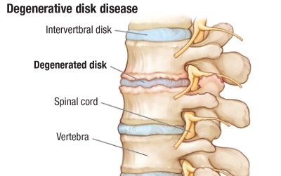
This article examines three major entities linked to lumbar DDD: lumbar spinal stenosis with neurogenic claudication, lumbar radiculopathy from disk herniations causing leg pain, and lumbar spondylolisthesis that results in bag and leg pain. For each entity we will discuss signs and symptoms of the three conditions, the diagnostic imaging used in diagnosing the diseased states, and management of these problems using both operative and non-operative strategies.
Lumbar Spinal Stenosis with Neurogenic Claudication
Lumbar spinal stenosis involves narrowing of the spinal canal, lateral recesses, or neural foramina, often due to DDD-related disk bulging, facet hypertrophy, ligamentum flavum thickening, and osteophyte formation. Neurogenic claudication (pseudoclaudication) arises when nerve roots or the cauda equina experience intermittent ischemia or compression during activity with the spine under a weight bearing load.
Signs and Symptoms:
Patients often report insidious onset of low back pain with radiating leg discomfort. Classic neurogenic claudication features bilateral (or unilateral) buttock, thigh, or calf pain, cramping, numbness, tingling, or weakness triggered by standing or walking-especially upright posture or lumbar extension. Symptoms typically relieve with sitting, forward flexion (e.g., leaning on a shopping cart-“shopping cart sign”), or lying down in a fetal position. Unlike vascular claudication, relief comes from posture change rather than just stopping, and patients can often cycle or walk uphill better than on flat ground. Severe cases may involve gait instability, foot drop, or bowel/bladder dysfunction. Back pain may be mechanical but is often less prominent than leg symptoms.
Imaging Modalities:
Plain radiographs (X-rays) assess alignment and are often performed with flexion-extension views to detect dynamic instability, disk height loss, spondylolisthesis, and osteophytes but lack soft tissue detail. MRI is the gold standard, visualizing central canal narrowing, thecal sac compression, nerve root impingement, and degenerative changes. CT or CT myelography serves as an alternative when MRI is contraindicated (e.g., pacemaker), offering superior bony detail for surgical planning.
Non-Operative Management:
Initial treatment is conservative for three to six months in mild-moderate cases. Physical therapy focuses on core strengthening, flexion-biased exercises, posture training, and stretching to reduce extension stress. NSAIDs, acetaminophen, or short-course muscle relaxants manage pain and inflammation. Epidural steroid injections provide targeted relief for radicular symptoms, often repeated as needed. Lifestyle modifications include weight loss, smoking cessation, and activity pacing. Bracing may help some patients.
Operative Management:
Surgery is indicated for refractory symptoms, progressive neurologic deficits, or cauda equina syndrome. Decompressive laminectomy (with or without foraminotomy) is common to relieve neural compression. For coexisting instability, instrumented fusion (e.g., posterolateral or interbody) may be added. Minimally invasive techniques reduce recovery time. Outcomes are generally good for leg pain relief, though back pain may persist. Risks include infection, instability, or adjacent segment degeneration.
Lumbar Radiculopathy from Lumbar Disk
Herniations Disk herniation occurs when the nucleus pulposus, (the gelatinous material in the center of the intervertebral disk), protrudes through the annulus fibrosus (the thick fibrotic wall that contains the disk material), often compressing the lumbar nerve roots. This is a frequent DDD sequela, especially at L4-L5 or L5-S1, causing sciatica or radiculopathy.
Signs and Symptoms:
Sudden or gradual onset of leg pain typically occurs follows lifting, bending, or trauma, though it can be spontaneous.
Key features include radicular leg pain (sharp, shooting, electric) worse than back pain, following a typical pattern on the skin (e.g., L5: lateral leg/dorsum of foot; S1: posterior calf/lateral foot). Pain intensifies with sitting, coughing, sneezing, or bearing down when using the bathroom. Neurologic deficits may include sensory loss, muscle weakness (e.g., foot drop in L5), or diminished reflexes. Positive straight-leg raise on examination reproduces these symptoms of radiating leg pain.
Severe, large, central herniations place the patient at risk for cauda equina syndrome (saddle anesthesia, bowel/bladder dysfunction, bilateral weakness)—a surgical emergency.
Imaging Modalities:
MRI (without contrast initially) is preferred for evaluation of all lumbar disk herniations, showing the herniation location (central, paracentral, foraminal), the compression of the nerve root and disk material characteristics. It differentiates from other causes like tumors or abscesses. CT myelography is an alternative for MRI-ineligible patients. Plain X-rays rule out other bony issues but miss soft disk pathology. Electrodiagnostic studies (EMG/NCS) confirm radiculopathy or rule out peripheral neuropathy when imaging is equivocal.
Non-Operative Management:
Most lumbar disk herniations (80-90%) resolve within six to twelve weeks. Activity modification (avoid prolonged sitting/bending), NSAIDs, oral steroids (short course), gabapentinoids for neuropathic pain, and physical therapy (McKenzie extension exercises, core stabilization) are first-line treatment modalities. Epidural steroid injections offer diagnostic and therapeutic value. Patients should stay active rather than bed rest.
Operative Management:
Microdiscectomy is the gold standard for persistent severe pain (>six weeks), progressive weakness, or cauda equina. It involves removing the herniated fragment via a small laminotomy. Endoscopic or minimally invasive variants minimize tissue disruption and are preferrable to open techniques. Fusion is rarely needed unless significant instability exists. Surgery provides faster pain relief but similar long-term outcomes to conservative care in many cases. Recurrence risk for disk herniations at the same level is ~5- 15%.
Lumbar Spondylolisthesis
Spondylolisthesis is anterior slippage of one vertebra over another, often degenerative (from DDD and facet arthritis, common at L4-L5 in older adults) or isthmic (pars defect, more common in younger patients).
Signs and Symptoms:
Many cases are asymptomatic. Symptomatic patients report mechanical low back pain worsened by extension/activity and relieved by rest/flexion. Neurogenic claudication or radiculopathy (often L5 root at L4-5 slip) may occur from associated stenosis. Hamstring tightness is common. High-grade slips (>50%) can cause visible deformity, gait changes, or cauda equina symptoms with pain that is often positional.
Imaging Modalities:
Lateral X-rays (standing flexion-extension) are initial and best for grading slippage and detecting instability. MRI evaluates nerve compression, disk degeneration, and stenosis. CT provides bony detail for pars defects or surgical planning. Bone scans may be used to detect acute stress often used in pediatric patients.
Non-Operative Management:
First-line treatment for low-grade (Grade I or II) slips without deficits: physical therapy (core/hamstring strengthening, flexion exercises), NSAIDs, activity modification, and bracing (temporary). Epidural injections address radicular pain. Weight management and smoking cessation aid outcomes.
Operative Management:
Indicated for high-grade slips, progressive slippage, refractory pain (>six months), or neurologic deficits. Decompression (laminectomy) plus instrumented fusion (often with interbody support) is standard to stabilize the segment. Minimally invasive options to treat high grade spondylolisthesis and demonstrate favorable outcomes for pain and function. Adjacent segment issues can arise long-term from either a traditional open technique or a minimally invasive one.
Conclusion and Broader Considerations
Lumbar DDD represents a spectrum of disabling mechanical and neurologic conditions. Multidisciplinary care-emphasizing patient education, lifestyle changes, and operative versus non-operative interventions-yields the best results. While most patients improve non-operatively, timely surgery prevents chronic disability in select cases. Emerging therapies like regenerative injections or advanced motion-preserving implants continue to evolve but are not widely available. For patients who ultimately require operative interventions, motion preservation techniques, minimally invasive and endoscopic interventions should strongly be considered compared to traditional open methods when appropriate.
Individuals should consult spine specialists for personalized evaluation, as imaging findings must correlate with clinical symptoms. My surgical philosophy is to provide the least amount of surgery necessary to get the patient the maximum amount of benefit from their degenerative lumbar spinal condition.
 Dr. Phillip Parry is a board-certified Neurosurgeon who joined Wellstar Health System in July of 2018 after having completed his military service with the US Air Force. Prior to his military service, Dr. Parry graduated from Tulane University School of Medicine in 2008 and completed residency at the University of Pittsburgh Medical Center from 2008-2015. He has completed fellowships in Endovascular Neurosurgery and minimally and maximally invasive spine surgery. He is currently the medical director for Spine at Wellstar and maintains a special interest the treatment of complex spinal disease.


